
Example 2
Example Instructions
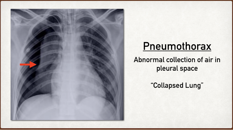
Slide 1
A pneumothorax is colloquially referred to as a "collapsed lung." Pneumothorax is created when the visceral and parietal pleura of a lung 🫁 are no longer are attached. Trauma, blebs (when there's too much coughing for example) or other injuries can all result in pneumothoraces. In this contest, you will be tracing pneumothoraces.
Slide 2
A pneumothorax is air that fills the newly created space between the visceral and parietal pleura. Look for the lack of lung lining or darker areas to make your annotation.
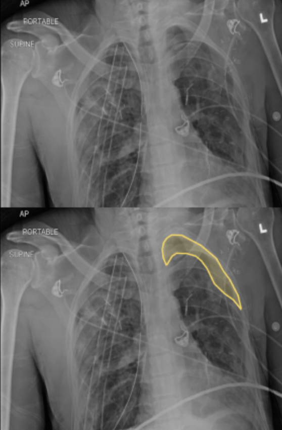
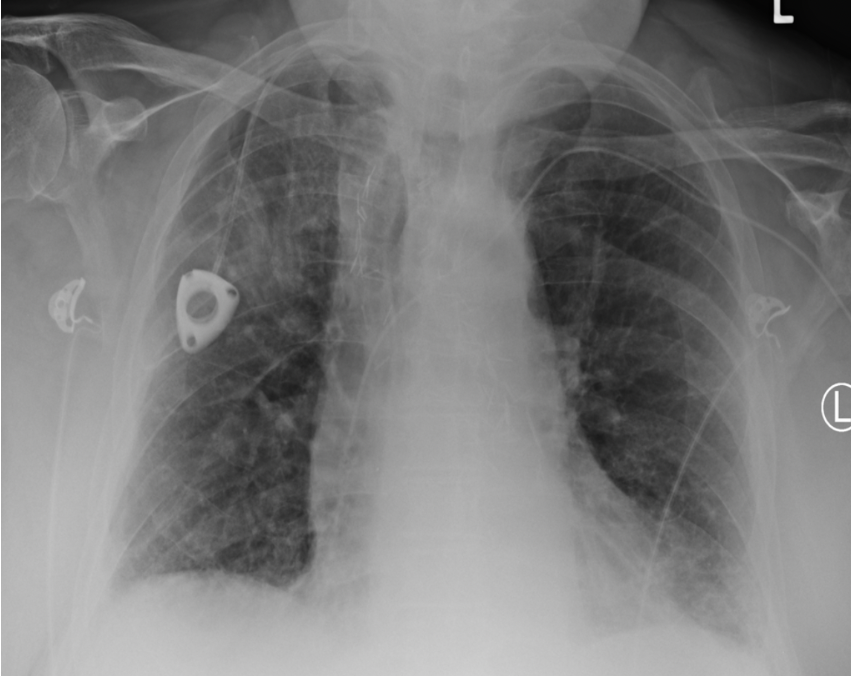
Slide 3
Here we can see on the top left side (anatomically) that we have a new area. This is causing a lack of lung lining in this location.
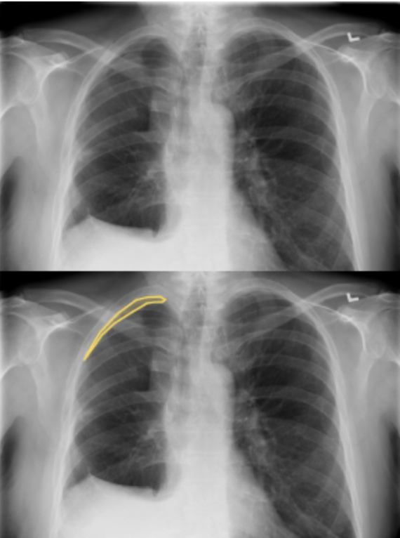
Slide 4
A pneumothorax is always a medical emergency until otherwise proven, as severe cases can be significantly life-threatening. Here is another example where the pneumothorax is in the top right (anatomically).
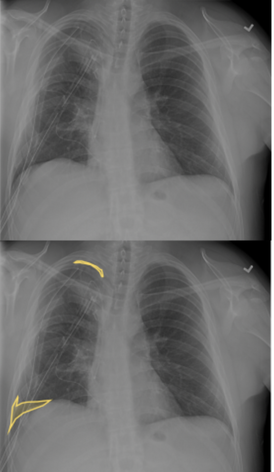
Slide 5
Some x-rays may show multiple areas of pneumothorax. Here we can see two different ones: at the top right and bottom right (anatomically).
Slide 6
Sometimes, there won't be any findings in these x-rays. Don't read in between the lines! Mark an x-ray without PNX present as "No findings."
Slide 7
⏰ Time to practice! The Practice section contains real cases that you will see in the competition. A pneumothorax is one of the hardest findings to discern, but your skills reading x-rays may save a life someday!